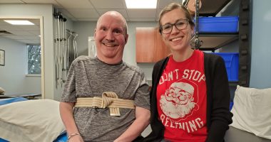
EMG helps track progress for SMA patients in physical therapy
Test reveals muscle activation at rest and during contractions
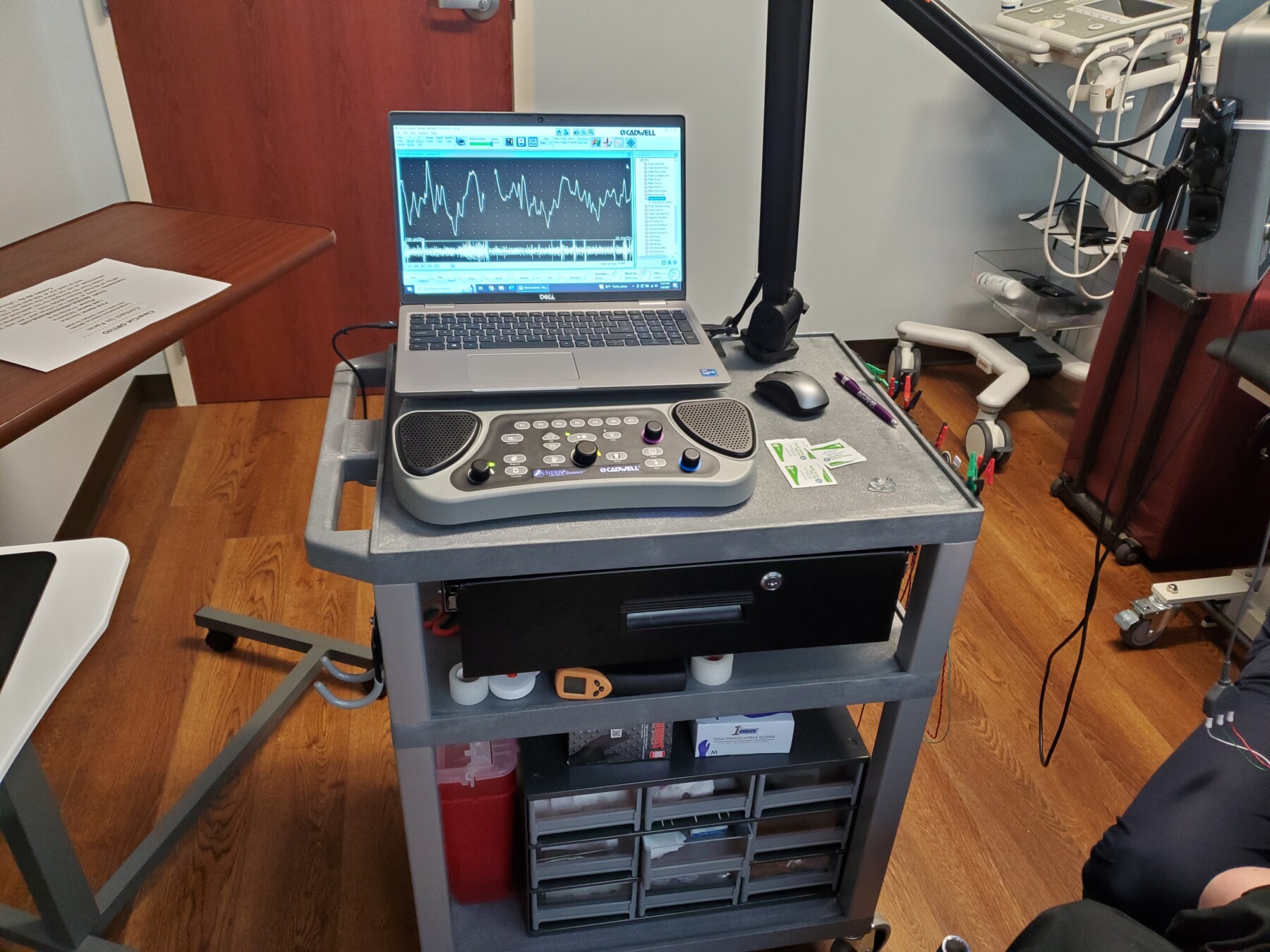
Electromyography (EMG), used to measure electrical activity in muscles. (Photo by Emily Jones)
Electromyography (EMG) is a test done with electrodes and/or needles inserted into or onto specific muscles to assess muscle activation at rest and during contraction. For Michael, my patient who has spinal muscular atrophy (SMA), we performed an EMG on different muscle groups to assess resting and activation contractions.
These muscle groups included anterior tibialis, which brings the toes up toward the face; bicep, which flexes the elbow; pronator group, which turns the hand toward the ground; and upper trapezius, which shrugs the shoulder up. All of these tests were conducted on the right side of Michael’s body.
In the anterior tibialis, we couldn’t get a solid reading due to increased swelling in his ankles and feet interrupting the signal. In all the other areas, we were able to see contractions with isometric movements with needle places in the muscle as well as reference electrodes. In the upper extremities, the right side was utilized due to that being the side Michael has the most activation.
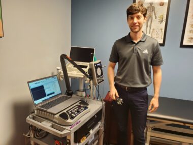
Ken Wheeler, the owner of ClearCut ORTHO in Fort Worth, Texas, uses electromyography to check electrical activity in muscles. (Photo by Emily Jones)
Scholar Rock is currently conducting a phase 3 clinical trial, called SAPPHIRE (NCT05156320), of their new muscle-targeted therapy apitegromab, and this clinical trial is scheduled to conclude in the third or fourth quarter of 2024. Since Scholar Rock has already been awarded fast track status by the U.S. Food and Drug Administration (FDA), they are hopeful apitegromab will be granted FDA approval before mid-2025. Fast track designation is intended to accelerate a therapy’s development and speed its approval by providing more frequent meetings with the FDA and discussions about the development plan.
Apitegromab will work by blocking the latent form of myostatin and will begin to allow strength building in specific muscle groups. We will then be able to track different objective measures, one being muscle recruitment with EMG readings. With traditional manual muscle testing, we use different movements of the body with manual resistance to monitor progress in strength throughout a patient’s program, but this isn’t possible with Michael due to his lack of active full range of motion. Now with EMG availability in our clinic, we are able to track these activations more closely with reduced range of motion available.
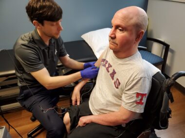
Ken Wheeler conducts an EMG on Michael Morale. (Photo by Emily Jones)
There are many reasons to utilize EMG assessments in the population of patients with SMA. Even though we are hoping to utilize EMG to measure an increase in muscle activation once apitegromab is released and Michael is approved for treatment, it could also be used to track the disease progression as muscle recruitment declines. Through the utilization of EMG technology and testing, we will have the functionality to be very specific in the muscle groups we target for movements that will increase Michael’s independence and improve his function.
Michael’s first goal is to independently drink from a cup without using a straw. Being able to accomplish this may seem minuscule to some, but this would greatly benefit Michael in that he would be able to freely drink water and other liquids as much as desired without having to ask for assistance.
Over the next two years, we will perform another EMG when we are closer to beginning apitegromab and will have a good baseline. Once that begins, we will discuss how frequently the EMG will be monitored depending on how we see how Michael is progressing. We are excited to utilize this technology to further increase Michael’s independence in a more efficient and effective way.
Patient perspective
My first EMG was performed in 1970 when I was 5 years old, and I remember this being an extremely traumatic experience. But then again, any 5 year old would feel the same when they saw doctors holding a handful of needles and learning these doctors were about to insert all of those needles in various parts of their body.
My second EMG was performed 20 years later in 1990. Within that 20-year period, needles became much smaller and my fear of them decreased significantly due to the number of needles that had been used on me. Few people relish the thought of having needles inserted into their muscles, but as those of us with SMA get older, we learn knowledge is much more important than the slight discomfort of the needles.
I feel this article is extremely important for physical therapists working with SMA patients. As new muscle-targeted therapies are developed over the coming years, doctors and patients need to utilize methods like EMG so they can better judge positive and negative reactions, especially with electrical activity within various muscle groups. Those of us with SMA have seen little improvement with regards to muscle mass and muscle strength while on our current treatments, but as these new muscle-targeted therapies are developed, EMG will be one of the tools that we use.


